Інші зображення цього випадку
Image
Isolated Posterior Cruciate Ligament Tear
- Image ID
- MPX2594_synpic20735
- Case U_id
- MPX2594
- Modality
- MR · MR - T2 weighted
- Plane
- Sagittal
- Location
- Musculoskeletal (Spine and Muscles)
- Age / Sex
- 34 / male
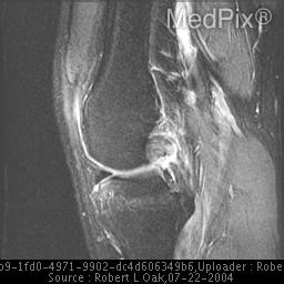
- Caption
- Increased signal within the anterior lateral tibial plateau concerning for an occult tibial plateau fracture. Note: image is fat suppressed.
- ACR Codes
- 4.4
Clinical case
- History
- Patient is status post motor vehicle injury. He complains of difficulty walking and knee pain.
- Exam
- The physical examination noted a joint effusion.
- Findings
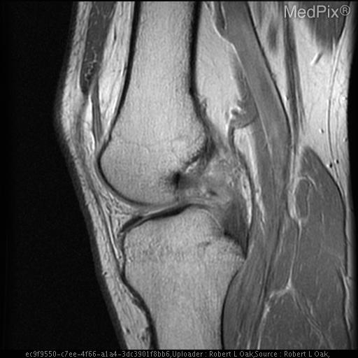
- MR images of the left knee demonstrate a large joint effusion. Heterogeneous high signal is seen within the posterior cruciate ligament, consistent with a complete tear. There is a significant degree of bone marrow edema within the anterior lateral tibial plateau, and an occult tibial plateau fracture cannot be completely excluded. Additionally, there is abnormal vertical high signal within the medical meniscus, consistent with a free edge traumatic tear. The lateral meniscus is normal. The medial and lateral collateral ligament complexes are normal. The anterior cruciate ligament is also normal.
- Differential Diagnosis
- A mid-substance interstitial lesion (most common). Complete disruption at the genu Avulsion at the tibial attachment (least common). - this requires reduction of osseus fragments to increase the chance of healing.
- Case Diagnosis
- Isolated Posterior Cruciate Ligament Tear
- Diagnosis By
- Surgically.
Topic
- Category
- Trauma
- ACR Code
- 4.4
Disease discussion
Posterior Cruciate Ligament Tear
PCL has it origin along the lateral aspect of medial femoral condyle and its attachement at the posterior intercondyloid fossa of the tibia. Both the PCL and ACL are intraarticular but extrasynovial
The PCL is a central stabilizer of the knee. It is composed of an anterolateral band and posteromedial band that tighten on flexion & extension respectively and restrict posterior tibial displacement on the femur. PCL is twice as strong as the ACL with a higher tensile strength and a larger cross sectional area. Therefore, the ACL is more commonly injured than the PCL. Injuries to PCL account for 5 – 20 % of knee ligament injuries and are usually associated with ACL, meniscus or collateral ligament damage. The most common location for PCL tear is the midportion representing 76% of all PCL injuries. Avulsion from the femur (36 – 55%) and from the tibia ( 22 – 42%) are the other two locations of PCL tear. The mechanism of injury is excessive rotation, hyperextension, dislocation or direct trauma while the knee is flexed. The most common causes of PCL injury are dashboard strikes in MVAs and contact sports. Clinically, the posterior drawer sign will be positive in 60% of PCL injuries.
Treatment for PCL tears include surgical repair and non-operative treatment. Isolated PCL tears are usually treated non-operatively. Surgical repair is usually reserved for symptomatic chronic PCL injury, acute bony avulsion, and combination injuries.