Інші зображення цього випадку
Image
Cuneiform Stress Fracture
- Image ID
- MPX2586_synpic17000
- Case U_id
- MPX2586
- Modality
- MR · MR - T2 weighted
- Plane
- Sagittal
- Location
- Musculoskeletal (Spine and Muscles)
- Age / Sex
- 78 / female
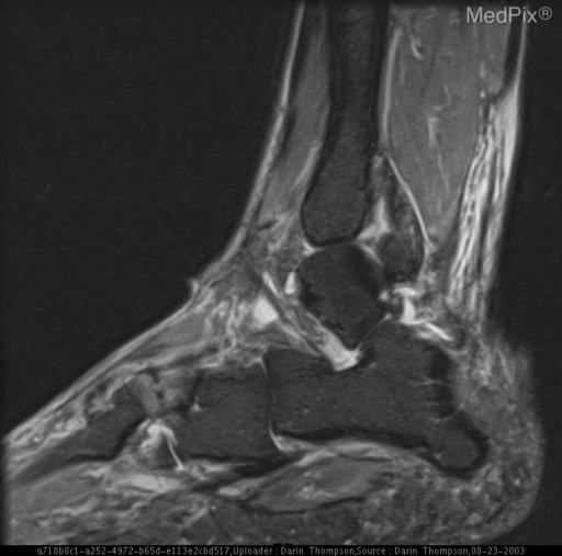
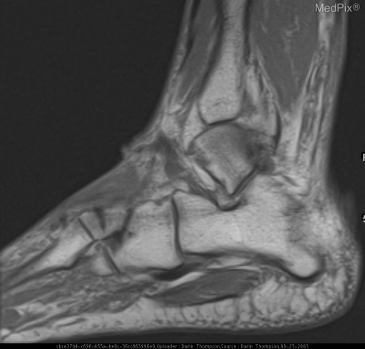
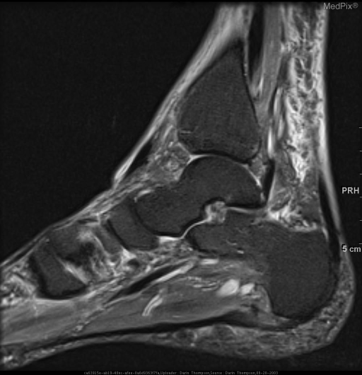
- Caption
- Bone marrow edema within the lateral cuneiform bone with transverse fracture line.
- ACR Codes
- 4.4
Clinical case
- History
- Foot pain. History of plantar fasciitis.
- Findings
- MRI: Bone marrow edema within the lateral cuneiform bone with transverse fracture line. Thickened plantar fascia with high T2 signal near calcaneal insertion also was seen consistent with plantar fasciitis.
- Differential Diagnosis
- -stress fracture -insufficiency fracture
- Case Diagnosis
- Cuneiform Stress Fracture
Topic
- Category
- Trauma
- ACR Code
- 4.4
Disease discussion
Stress fractures of the cuneiform bones are rare. More typical types of stress fractures include metatarsal ("march" fractures) and in the lower extremities in athletes, joggers, and dancers. Common sites include the calcaneus or other tarsal bones (ie navicular, less commonly), fibula, tibia, femur, metatarsal, pelvis, upper extremity and ribs.
Stress fractures can occur in normal or abnormal bones subjected to chronic loading. Resnick describes two types of stress fractures. 1. Fatigue fracture from abnormal stress to normal bone. and 2. Insuffieciency fracture, with normal stress on abnormal bone.
Causes include RA, osteoporosis, Paget's, osteomalacia, renal osteodystrophy, and radiation.
Plain film plays an essential role in stress fracture diagnosis; however, bone scan and MRI have better diagnostic sensitivity. MRI has comparable sensitivity and specificity superior to bone scan.
Stress fractures appear most typically as a linear zone of low signal on T1WI and a linear area of low SI surrounded by broader high SI on T2WI. Prompt diagnosis and treatment is key in preventing a tarsal stress fracture from becoming a chronic source of foot pain.