Інші зображення цього випадку
Image
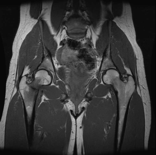
Stress fracture right femoral neck.
- Image ID
- MPX2502_synpic24909
- Case U_id
- MPX2502
- Modality
- MR · MR - T1W - noncontrast
- Plane
- Coronal
- Location
- Musculoskeletal (Spine and Muscles)
- Age / Sex
- 21 / female
- Caption
- Coronal T1-weighted MR image of the hips shows linear hypointensity of femoral neck with surrounding low-signal-intensity bone marrow edema
- ACR Codes
- 4.4
Clinical case
- History
- 21 year old female with right hip pain after recent increase in physical activity.
- Exam
- Patient’s chart unavailable for review
- Findings
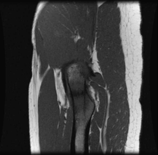
- A-P radiograph of the hips shows no apparent fracture Follow-up A-P radiograph of the hips shows a sclerotic line in inferior aspect of the femoral neck consistent with a healing stress fracture. MRI- T1 weighted images showing linear hypointensity at the inferior aspect of the right femoral neck with consistent with a stress fracture. This area became high signal intensity on STIR MR images.
- Differential Diagnosis
- Stress fracture right femoral neck
- Case Diagnosis
- Stress fracture right femoral neck.
Topic
- Category
- Trauma
- ACR Code
- 4.4
Disease discussion
Stress fractures occur as a result of repetitive submaximal stress on normal bone, which results in a region of bone undergoing accelerated bone remodeling. This in turn can lead to microtrabecular breaks and ultimate cortical injury as the rate of osteoclast activity exceeds the rate of new bone formation. Two types of femoral neck stress fractures have been identified in adolescents/young adults-transverse fractures to the superior portion of the femoral neck, and compression fractures to the inferior portion of the femoral neck. Management of the more proximal fracture is with closed reduction and internal fixation. Initially, conservative therapy (non-weight bearing, reduction of physical activity) can be attempted for stress fractures involving the distal femoral neck, but failure of conservative therapy or delay in diagnosis will also lead to surgical intervention.