Image
Chance Fracture T11
- Image ID
- MPX2454_synpic39850
- Case U_id
- MPX2454
- Modality
- MR · MR - Other Pulse Seq.
- Plane
- Sagittal
- Location
- Spine (Spine and Muscles)
- Age / Sex
- 23 / male
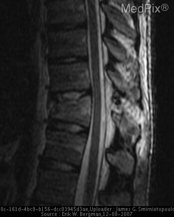
- Caption
- MRI (Sagital STIR): Increased signal through T11 body and posterior elements secondary to fracture. The important findings here are: minimal posterior distortion of the fracture vertebral body into spinal canal without evidence of cord compromise or compression. No abnormal cord signal. (Close-Up)
- ACR Codes
- 0.0
Clinical case
- History
- 23 y.o. man after a high fall
- Exam
- Multiple trauma
- Findings
- AP/LAT T-SPINE: Anterior compression fracture of T11 with loss of approximately 50% of height. AP view demonstrates abnormal pedicles with linear fractures and widening bilaterally. Posterior elements not well seen on lateral secondary to spine board. CT (Sag recon): Horizontal fracture of T11 vertebral body extending horizontally throught posterior elements MRI (Sag ): No cord compression or abnormal signal
- Differential Diagnosis
- The differential diagnosis initially after plain films alone include (compression, burst, chance and fracture dislocation). The abnormal appearance of the pedicles with horizontal lucent fractures and widening rules-out compression fracture alone. The involvement of the posterior elements and non-comminution of body fracture rules-out a burst fracture. The fact that there are no associated dislocations rules-out fracture dislocation.
- Case Diagnosis
- Chance Fracture T11
- Diagnosis By
- CT sagittal reconstruction
Topic
- Category
- Trauma
- ACR Code
- 4.4
Disease discussion
There are 4 types of thoracolumbar fractures often described. These include compression, burst, Chance ("seatbelt"), and fracture-dislocation.
Compression fractures show anterior column compression (anterior vertebral body) with usually no middle or post column involvement. Burst fractures produce anterior and middle column compression (vertebral body and anterior neural arch). Fracture-dislocations involve all three (anterior, middle, and posterior) columns which results in intervertebral subluxation or dislocation.
Chance or Chance-type fractures involve the posterior, middle, and occasionally the anterior columns. The injury results from a severe flexion of the spine with the fulcrum not being the the anterior vertebral body as in compression fractures. Instead, the anterior abdominal wall (i.e. where the lap seatbelt fits) is the fulcrum area with hyperflexion at thoraco-lumbar junction (e.g. L1). This creates a distraction force on the posterior and middle columns of the vertebra, creating a horizontal fracture extending posterior to middle. This fracture may then extend anteriorly throught vertebral body (with an assoc compression of anterior body).
Chance fracture is an “eponym”, named after G.Q. Chance who described this injury in 1948 as anterior wedging, compression, and a frature through the body, lamina, and spinous process. Because of the mechanisms of injury (flexion over a seat belt) about ½ are associated with intra-abdominal trauma.
Burst and fracture dislocations have a high incidence of instability. Chance fractures also have potential for instability although not as likely as the former two. Since radiographs may not clearly demonstrate the exact plane and extent of this fracture, CT (with sagital reconstruction is quite useful). Axial spine CT alone often won't delineate the fracture well due the fracture's horizontal (axial) nature. MRI can help determine cord compression and injury.