Інші зображення цього випадку
Image
Epidural Hematoma of middle fossa (temporal bone)
- Image ID
- MPX2427_synpic4861
- Case U_id
- MPX2427
- Modality
- CT · CT - noncontrast
- Plane
- Axial
- Location
- Brain and Neuro (Head)
- Age / Sex
- 5 / male
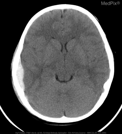
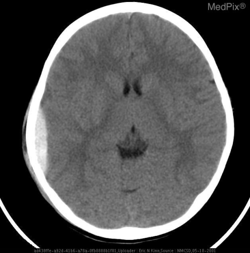
- Caption
- Rt temporal-occipital epidural hematoma with mild mass effect on right lateral ventricle.
- ACR Codes
- -1.-1
Clinical case
- History
- 5 year old male S/P fall from shopping cart. Now presents with lethargy and headache.
- Findings
- Rt temporal-occipital epidural hematoma with mild mass effect on right lateral ventricle.
- Case Diagnosis
- Epidural Hematoma of middle fossa (temporal bone)
Topic
- Category
- Trauma
- ACR Code
- 1.4
Disease discussion
Watch Video - http://youtu.be/30XPVg0DWH4
An epidural hematoma (EDH) is an accumulation of blood between the outer (periosteal) layer of the dura mater (literally "tough mother") and the naked bone of the inner table of the skull. Most patients will have an acute clinical presentation. The blood will, therefore, be hyperdense (white) on a plain CT scan. The most common source of bleeding is a laceration or tear in one of the meningeal arteries that feed the dura itself. Posterior or occipital epidurals may develop from lacerations of the dural transverse sinus.
More than 90% of patients with an EDH will have a skull fracture (and associated scalp trauma) in addition to the EDH. The temporal bone and the underlying middle cranial fossa are the most commonly affected areas. Because of the firm fixation of the dura to the suture, the hematoma only rarely crosses a cranial suture.
Some patients are knocked unconscious by the initial head impact (a "concussion") - but then wake up and may appear normal. This is often described as the "lucid interval" and occurs in about 40% of patients with epidural hematoma.
Dr. Paul Cooper, in his book "Head Injury", reports these five patterns of the clinical EDH:(U=unconscious; L=lucid; C=conscious)
1. U-->L-->U: 11 to 67%
2. U throughout: 19 to 56%
3. U-->C: 16 to 26%
4. C-->U: 7 to 21%
5. C throughout: 9 to 25%
6. unknown or unstated: 5%
NOTE: A significant number of patients do not even have a transient loss of consciousness. (Data courtesy of Vern Armbrustmacher).
As the blood accumulates from bleeding meningeal arteries, it forms a bi-convex or lens shaped mass that pushes the brain inward and downward - causing brain herniation.
The acute formation of the EDH often causes secondary downward transtentorial brain herniation. This, in turn, may cause neurologic signs,including oculomotor nerve (CNN-3) dysfunction and a "blown pupil" (fixed and dilated).
Many patients have the injury, but appear to be alright afterwards. As the blood accumulates and the brain begins to shift, they may slip into a coma. This is "lucid interval" is also sometimes described as "Talk and Die". Sadly, this is not rare, and in 2009 Tony Award winning actress Natasha Richardson died from an epidural hematoma. She had a lucid interval after falling while skiing, not wearing a helmet.
• http://www.sciam.com/article.cfm?id=talk-and-die-richardson
• http://abcnews.go.com/Entertainment/Movies/story?id=7119825&page=1'>http://abcnews.go.com/Entertainment/Movies/story?id=7119825&page=1
• http://www.cnn.com/2009/HEALTH/03/18/brain.injury/index.html?iref=mpstoryview
• http://www.washingtonpost.com/wp-dyn/content/article/2009/03/19/AR2009031902515.html
• http://www.baltimoresun.com/news/health/bal-epidural-hematoma-0318,0,7024060.story
• http://abcnews.go.com/Entertainment/Movies/story?id=7119825&page=1
For more on Intracranial Hemorrhage: http://rad.usuhs.mil/rad/handouts/jsmirnio/Intracranial_Hemorrhage_%20RSNA_2008_HO.pdf
https://www.youtube.com/watch?v=30XPVg0DWH4
https://www.youtube.com/watch?v=jiscvATspCA