Інші зображення цього випадку
Image
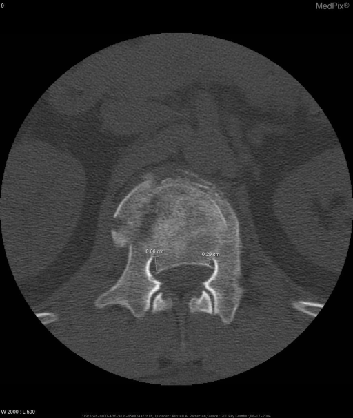
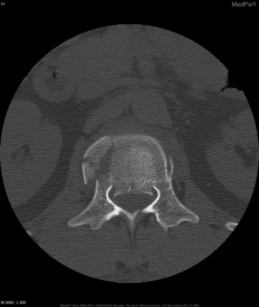
Diagnosis: L1 Burst Fracture.
- Image ID
- MPX2354_synpic20936
- Case U_id
- MPX2354
- Modality
- CT · CT - noncontrast
- Plane
- Axial
- Location
- Spine (Spine and Muscles)
- Age / Sex
- 34 / male
- Caption
- CT Lumbar spine
- ACR Codes
- 3.9
Clinical case
- History
- History: 34 y/o active duty white male with 2/10 lower back pain after a fall. The fall occurred while exiting a helicopter at an unknown height by “fast-roping:” sliding down a rope suspended from the helicopter using only hands and feet to control speed of descent. Initially he landed on his feet, but then he fell onto his sacrum. The onset of pain was immediate and localized to his lower back. Since the date of injury, the patient denies change in quality or radiation of pain, lower extremity numbness or weakness, loss of bladder or bowel control, erectile dysfunction.
- Exam
- Physical Exam and Laboratory: Pertinent physical exam findings: Lower extremities were neurovascularly intact distally with brisk capillary refill and no edema. Patellar and Achilles deep tendon reflexes were 2+ and symmetric. Babinksi sign was negative. Strength was 5/5 in bilateral iliopsoas, quadriceps, hamstrings, tibialis anterior, gastrocnemius, extensor hallicus longus. Cranial nerves II – XII were intact. Sensation and proprioception were normal and symmetric in both upper and lower extremities. There was no tenderness to palpation on the patients back or costovertebral angle tenderness. Gait was normal.
- Findings
- Imaging Findings: L-Spine: Lateral view shows anterior wedging of the L1 vertebral body. The superior end-plate of L1 is displaced posteriorly approximately 1 cm relative to the inferior end plate of T12. The inferior end-plate of L1 is displaced posteriorly approximately 0.5 cm relative to the superior end-plate of L2. CT: Fracture of the L1 vertebral body with posterior displacement of the central fragment, right lateral displacement of the right fragment.
- Differential Diagnosis
- Differential Diagnosis for these findings in this case: L1 Burst Fracture
- Case Diagnosis
- Diagnosis: L1 Burst Fracture.
Topic
- Category
- Trauma
- ACR Code
- 3.4
Disease discussion
Burst fractures of the lumbar spine occur when the anterior (anterior longitudinal ligament, anterior half of the vertebral body, and anterior portion of the annulus fibrosis) and middle (posterior longitudinal ligament, posterior half of the veterbral body, and posterior portion of the annulus fibrosis) columns of the spine fail under a compressive load. This forces the intervertebral disc into the vertebral end-plate, causing the vertebral body to burst - with outward displacement of its fragments. (Canale: Campbell’s Operative Orthopaedics, 10th ed., 2003. pp1643-4)
Patients should first be evaluated with radiographs. To determine the position and extent of fracture fragments, CT should be performed. If patients have symptoms of spinal cord compression, MRI should be performed.
Although conservative treatment of a burst fracture can be attempted using a brace, surgical correction is indicated if a posteriorly displaced fragment may cause neurological symptoms. Pedicle screws with rods can be used to fix the vertebral bodies cephalad and caudal to the burst vertebra. Titanium cages with bone graft can be left in place after corpectomy of the fractured body to promote fusion and rotational stability. (Mariotti AJ, Diwan AD, Current concepts in anterior surgery for thoracolumbar trauma. Ortho Clin North Am 2002;33(2):403-12)
Plain film radiographs and CT can be used to assess postoperative hardware stability. Physical exam will also ensure that neurological symptoms have not developed.