Інші зображення цього випадку
Image
Meniscal Tear (radiologic and athroscopic confirmation)
- Image ID
- MPX2316_synpic12143
- Case U_id
- MPX2316
- Modality
- MR · MR - T2 weighted
- Plane
- Coronal
- Location
- Musculoskeletal (Spine and Muscles)
- Age / Sex
- 30 / male
- Caption
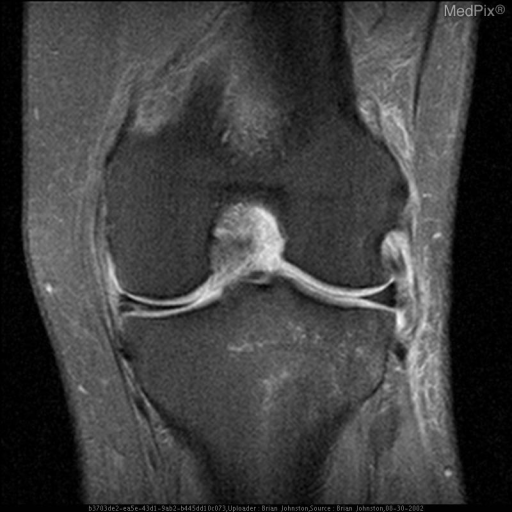
- 1)1 This is a T2 weighted coronal image of the Left knee with fat saturation showing abnormal signal in the medial meniscus which clearly contacts the articular surface. This is consistent with a meniscal tear. Also note edema surrounding the lateral collateral ligament (sprain)
- ACR Codes
- 4.4
Clinical case
- History
- 30 y.o. man with chronic left knee pain after an injury
- Exam
- No exam or lab findings available
- Findings
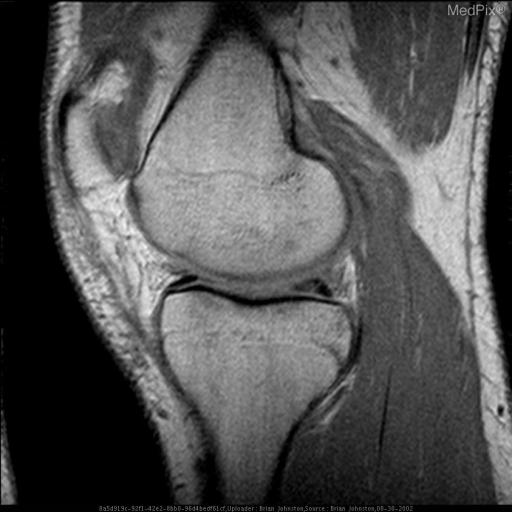
- 1) This is a T2 weighted coronal image of the Left knee with fat saturation showing abnormal signal in the medial meniscus which clearly contacts the articular surface. This is consistent with a meniscal tear. Also note edema surrounding the lateral collateral ligament (sprain). 2) This Sagittal proton density image of the left knee reveals linear abnormal signal in the posterior horn of the medial meniscus
- Differential Diagnosis
- Knee, medial meniscus tear
- Case Diagnosis
- Meniscal Tear (radiologic and athroscopic confirmation)
- Diagnosis By
- Imaging and arthroscopic surgery
Topic
- Category
- Trauma
- ACR Code
- -1.-1
Disease discussion
Meniscal tears are common injuries in both the sport and non-sport population with acutely torn menisci cases numbering at 61 per 100,000. (1) The medial meniscus is the more commonly torn than the lateral meniscus in sports injuries where the mechanism for injury “is a compressive, rotational, and shearing force.” (1)
MRI is reported to have a 95% accuracy of detection rate for meniscal tears, but studies note that this number also depends on magnetic field strength of the scanners. (1)
The key to diagnosing meniscal tears on MRI is by noting abnormal signal in the meniscus. Nevertheless, the abnormal signal must clearly touch the inferior or superior articular surface to call a tear on MRI. Note that a normal meniscus can have some signal within it. Tears that have minimal symptoms can be left without treatment, but oftentimes tears are treated at the time of arthroscopy with shaving or debridement.