Інші зображення цього випадку
Image
Anterior Cruciate Ligament Disruption
- Image ID
- MPX2300_synpic31227
- Case U_id
- MPX2300
- Modality
- MR · MR - T2 weighted
- Plane
- Sagittal
- Location
- Musculoskeletal (Spine and Muscles)
- Age / Sex
- 21 / male
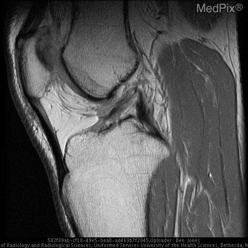
- Caption
- Sagittal T2-weighted image through the level of the intercondylar notch demonstrates disruption of the proximal fibers of the ACL.
- ACR Codes
- 4.4
Clinical case
- History
- The patient was a 21-year man, who twisted his knee while playing in a softball game. Following the injury, he complained of difficulty running, pain in the posterolateral aspect of the knee and joint swelling.
- Exam
- Initial physical examination of the knee revealed a small joint effusion, but no deformity. The knee demonstrated full range of motion and there was no documentation of instability.
- Findings
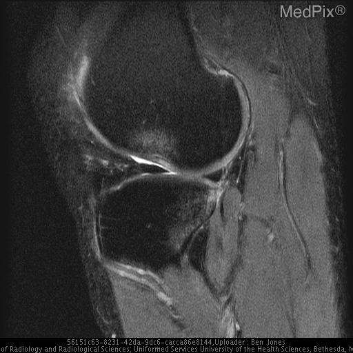
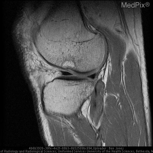
- Lateral radiograph of the knee demonstrates a deep, irregular appearing lateral femoral sulcus, and a small joint effusion, but is otherwise normal (Fig 1). Follow up MR imaging demonstrates marrow edema in the lateral femoral condyle and in the posterior tibial plateau. The lateral femoral sulcus appears deepened and irregular in contour. T2-weighted sagittal image through the region of the intercondylar notch demonstrates complete disruption of the anterior cruciate ligament (Fig 2).
- Differential Diagnosis
- Anterior Cruciate Ligament Disruption
- Case Diagnosis
- Anterior Cruciate Ligament Disruption
- Diagnosis By
- Follow up MRI confirmed the complete disruption of the anterior cruciate ligament.
Topic
- Category
- Trauma
- ACR Code
- 4.4
Disease discussion
Several apparently minor osseous abnormalities of the knee have been described on conventional radiographs that are associated with more serious but radiographically occult soft tissue injuries. These include avulsion fracture of the anterior tibial spine and avulsion fracture of the lateral tibial plateau (Segond fracture), both associated with ACL disruption and avulsion fracture of the posterior intercondylar insertion of the posterior cruciate ligament (2-4). The deep femoral sulcus sign, however, is the most common radiographic sign associated with ligamentous injury of the knee. Because it is an impaction injury rather than an avulsion injury, it can be a subtle finding on radiographs, and one that is often overlooked.
A normal lateral radiograph of the knee demonstrates a normal shallow groove along the lateral femoral condyle that is formed by the junction of the tibiofemoral and patellofemoral curvatures. This notch is referred to as the lateral condylopatellar sulcus and it is normally smooth and symmetric in appearance measuring less than 1.5 mm in depth (5). During the pivot shift mechanism of injury, the knee while in slight flexion undergoes a twisting injury and valgus stress that leads to disruption of the ACL thus allowing anterior translation of the tibia relative to the femur. As the tibia and femur return to their normal position, the lateral femoral condyle impacts the posterolateral tibial plateau resulting in an osteochondral impaction fracture of the lateral femoral condyle.
The radiograph will demonstrate an irregular asymmetric appearing notch that is greater than 2 mm in depth. This is referred to as the “deep lateral femoral sulcus” sign. The depth of the notch is measured by drawing a line tangential to the sulcus along the articular surface of the lateral femoral condyle and then by measuring the distance from the line to the depth of the sulcus (Fig 5). The presence of a deep lateral femoral sulcus is nearly always associated with ACL disruption but lacks sensitivity; the absence of the sign therefore does not exclude the presence of an ACL tear (5).
MR imaging also demonstrates an irregular appearing deep lateral femoral sulcus and in addition it will demonstrate marrow edema within the lateral femoral condyle and the posterior tibial plateau secondary to the impaction injury (6). This pattern of marrow edema has been referred to as the “pivot shift” marrow edema pattern and is also highly specific for ACL injury. See figures 2, 3 and 4 for examples in our case. MR imaging will demonstrate the ACL disruption along with associated injuries of internal derangement to include meniscal tear, chondral injury and collateral ligament tear.
The “deep lateral femoral sulcus” sign is a subtle radiographic finding that when present on a plain film can provide an important clue that an ACL injury has occurred. It results from an impaction of the lateral femoral condyle against the posterior tibial plateau at the time of ACL disruption. Identification of this finding on radiographs of the knee should prompt further evaluation of the knee with MR imaging.