Image
Atlas fracture
- Image ID
- MPX2258_synpic42061
- Case U_id
- MPX2258
- Modality
- CT · CT - noncontrast
- Plane
- Axial
- Location
- Spine (Spine and Muscles)
- Age / Sex
- 20 / male
- Caption
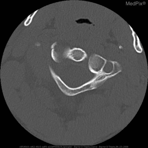
- Fracture of the Right lateral mass of the Atlas (C-1)
- ACR Codes
- 3.4
Clinical case
- History
- 20 y/o male ejected from a vehicle during a rollover crash. The patient reports a brief loss of consciousness.
- Exam
- His Glasgow Coma Score (GCS) was 15 when initially evaluated. Exam notable for multiple facial abrasions and contusions. There were no focal neurologic deficits.
- Findings
- Minimally displaced fracture of the Right lateral mass of the Atlas (first cervical vertabra).
- Differential Diagnosis
- • Atlas fracture • Transverse ligament injury
- Case Diagnosis
- Atlas fracture
- Diagnosis By
- computed tomography
Topic
- Category
- Trauma
- ACR Code
- 3.4
Disease discussion
Synonyms: Jefferson fracture, burst fracture
Associations/Predisposing Factors: motor vehicle crashes, sports injury (e.g. football), falls, violence
Common Locations: posterior arch, lateral mass, bilateral anterior and posterior arches (burst or Jefferson fracture).
Demographics: The majority of spinal fractures and dislocations occur in the cervical spine due to its mobility. The incidence is 25,000. Atlas fractures represent 10% or cervical spine injuries and 2% or all spine injuries. Most cervical spine injuries occur in males age 15-24.
Radiology: Atlas pathology can be detected by plain film images, especially with the odontoid, or open-mouth, view where lateral displacement of greater than 6.9mm is indicative of a fracture. However, this modality has low sensitivity so diagnosis is facilitated by computed tomography(CT). Magnetic resonance imagine (MRI) is indicated for investigating soft tissue injury, such as when ligamentous disruption is suspected or when there are neurologic deficits. Arteriography may be necessary to evaluate vascular injury.
Prognosis and Treatment: Isolated posterior arch and non- or minimally-displaced Jefferson fractures are considered stable. Treatment is a rigid cervical collar for 8-12 weeks with follow-up imaging to re-evaluate stability. The patient is expected to resume normal activity once the fracture has healed. Any fracture where the lateral mass is displaced 7mm or more is considered unstable. This requires halo traction for 3-6 weeks followed by further immobilization with a halo vest until stability is confirmed. Even with an unstable fracture, the prognosis is favorable provided that there are no other associated injures (such as neurologic insults).