Інші зображення цього випадку
Image
1. Mid-substance anterior cruciate ligament tear. 2. Lateral capsular avulsion consistent with Segond fracture. 3. Partial thickness tear of medial collateral ligament.
- Image ID
- MPX2097_synpic19231
- Case U_id
- MPX2097
- Modality
- MR · MR - T1W - noncontrast
- Plane
- Coronal
- Location
- Musculoskeletal (Spine and Muscles)
- Age / Sex
- 47 / female
- Caption
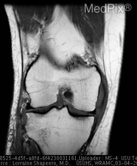
- T1-weighted coronal MR image shows indistinctness of the medial collateral ligament at the joint line and low-signal- intensity edema of the lateral tibial plateau and defect of the Segond fracture.
- ACR Codes
- 4.4
Clinical case
- History
- 47 y/o female status/post-skiing accident in which the patient’s right knee was clipped by a snowboarder while her leg was planted and internally rotated.
- Exam
- Tenderness and swelling of the right knee, + Lachman/anterior drawer, + valgus stress test. No laboratory test results were available.
- Findings
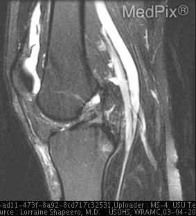
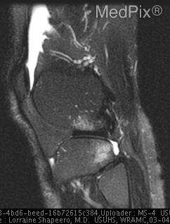
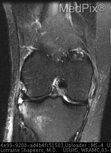
- 1. A-P radiograph of the knee taken at referring facility shows a Segond fracture. 2. FSE proton density with fat saturation sagittal oblique MR image shows a mid-substance anterior cruciate ligament tear with increased obliquity of the ACL and a joint effusion. Edema of posterior tibial plateau correlative with the Segond fracture is also seen. 3. More lateral FSE proton density with fat saturation sagittal oblique MR image shows both the high-signal-intensity edema of the Segond fracture and edema of the lateral femoral condyle, injuries seen with ACL tear. 4. T1-weighted coronal MR image shows indistinctness of the medial collateral ligament at the joint line and low-signal- intensity edema of the lateral tibial plateau and defect of the Segond fracture. 5. FSE T2-weighted with fat saturation coronal MR image shows the increased signal intensity within the medial collateral ligament consistent with a partial thickness tear. The Segond fracture is seen as a lateral capsule avulsion with a focal osseous deficit at the lateral proximal tibia. Associated with this is high-signal-intensity edema/contusion of the lateral tibial plateau and lateral femoral condyle.
- Differential Diagnosis
- Combined findings are characteristic for ACL, MCL tears and Segond fracture
- Case Diagnosis
- 1. Mid-substance anterior cruciate ligament tear. 2. Lateral capsular avulsion consistent with Segond fracture. 3. Partial thickness tear of medial collateral ligament.
Topic
- Category
- Trauma
- ACR Code
- 4.4
Disease discussion
A Segond fracture is a cortical avulsion of the tibia at the site of insertion of the lateral capsular ligament. This fracture results from excessive internal rotation and varus stress of the knee. Segond fractures are frequently associated with other internal knee derangements including ACL tear (75-100%), meniscal tears - particularly medial meniscus (66-75%), and other avulsion fractures of the fibular head or intercondylar eminence. Therefore, a patient with a Segond fracture, which is usually best seen on A/P radiographs, should be further evaluated by MRI for anterior cruciate ligament and other associated bone, ligamentous, and meniscal injuries. Sagittal T2W MRI with fat saturation is 95% sensitive and 98% specific for showing ACL tears. Treatment for Segond fracture is generally conservative for non-displaced/non-inverted fractures. Treatment for ACL tear is surgical for the athletic individual who wishes to return to his or her level of activity. Best results are usually achieved with a patellar tendon autograft.