Інші зображення цього випадку
Image
Osteochondritis dissecans.
- Image ID
- MPX2069_synpic24286
- Case U_id
- MPX2069
- Modality
- MR · MR - FLAIR
- Plane
- Coronal
- Location
- Musculoskeletal (Spine and Muscles)
- Age / Sex
- 35 / male
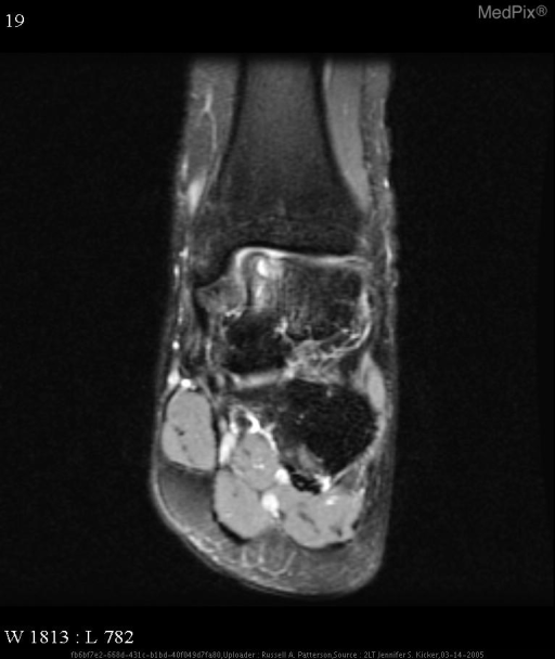
- Caption
- Coronal proton-density MR image with fat saturation shows a focal high-signal-intensity osteochondral lesion of the medial talar dome with associated edema in the talus.
- ACR Codes
- 4.4
Clinical case
- History
- Patient is a 35-year-old man with a history of a left distal fibular fracture in January 2003. He now presents with repeated episodes of pain and swelling around the left lateral malleolus after playing basketball.
- Exam
- Physical exam was unremarkable. Complete blood count and basic metabolic panel were within normal limits.
- Findings
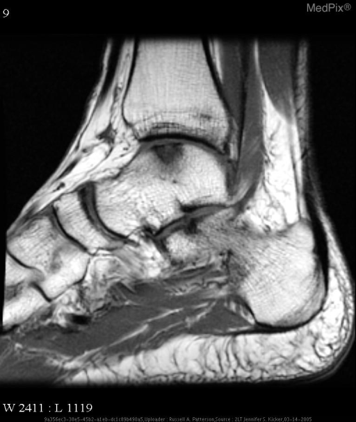
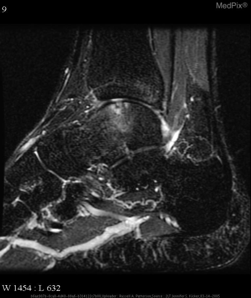
- AP and mortise radiographs of the left ankle shows a lucency in the medial talar dome. T1-weighted sagittal MR image of the left ankle shows a focus of low signal intensity in the talar dome. Coronal proton-density MR image with fat saturation and sagittal STIR MR image shows a focal high-signal-intensity osteochondral lesion of the medial talar dome with associated edema in the talus
- Differential Diagnosis
- 1.Osteochondral fracture 2. Osteochondritis dissecans
- Case Diagnosis
- Osteochondritis dissecans.
Topic
- Category
- Trauma
- ACR Code
- 4.4
Disease discussion
Osteochondritis dissecans (OCD) is a painful, usually unilateral condition commonly affecting individuals in the 20 to year old age group (men more frequently than women). In this lesion, a segment of articular cartilage and attached subchondral bone becomes partially or completely separated from the underlying parent bone. Etiology is thought to be secondary to trauma. Medial talar dome injury may be related to plantar flexion of the foot with accompanying inversion, followed by rotation of the tibia on the talus.
The use of MR imaging in the evaluation of OCD is credited in large part to the studies of De Smet. Specifically, he focused on the correlation between signal intensity and fragment stability. High signal intensity at the junction between the fragment and the parent bone on a T2 weighted image was a strong predictor of an unstable lesion.