Інші зображення цього випадку
Image
HILL-SACHS DEFORMITY WITH ASSOCIATED BANKART LESION
- Image ID
- MPX2043_synpic17061
- Case U_id
- MPX2043
- Modality
- MR · MR - PDW Proton Density
- Plane
- Axial
- Location
- Musculoskeletal (Spine and Muscles)
- Age / Sex
- 26 / male
- Caption
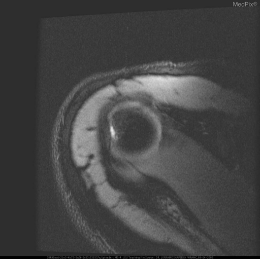
- FSE Proton Density W/ FAT SAT. AXIAL MR IMAGES OF RIGHT SHOULDER SHOW: ON THE MORE SUPERIOR IMAGE, FLATTENING OF THE POSTEROLATERAL ASPECT OF THE HUMERAL HEAD WITH HIGH SIGNAL INTENSITY IN THE HUMERAL HEAD AND OVERLYING CARTILAGE CONSISTENT WITH EDEMA/CONTUSION WITH HILL-SACH'S DEFORMITY.
- ACR Codes
- 4.4
Clinical case
- History
- H/O THREE ANTERIOR SHOULDER DISLOCATIONS SINCE AGE 21, ALL PLAYING RUGBY.
- Exam
- JOINT LAXITY ASSOCIATED WITH OVERHEAD AND CROSS-ARM MOVEMENT DIFFICULTY. OCCASIONAL LOCKING SYMPTOMS.
- Findings
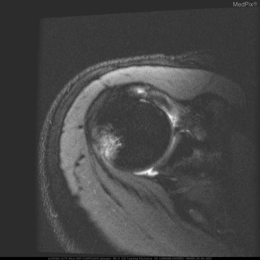
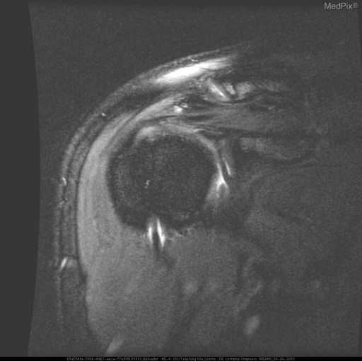
- A-P RADIOGRAPHS: SUBCHONDRAL SCLEROSIS IN THE REGION OF THE GLENOID TUBERCLE WITH DEPRESSION OF POSTERO-LATERAL HUMERAL HEAD ON EXTERNAL ROTATION. REMAINDER OF BONES NORMAL; NO BANKART LESION APPRECIATED. TWO FSE PROTON DENSITY W/ FAT SAT. AXIAL MR IMAGES OF RIGHT SHOULDER SHOW: 1. MORE SUPERIOR IMAGE, AT LEVEL OF SUPERIOR GLENOID, FLATTENING OF POSTERIOR-LATERAL ASPECT CONSISTENT WITH HILL-SACHS DEFORMITY. HIGH SIGNAL INTENSITY AROUND HUMERAL HEAD REPRESENTS CARTILAGINOUS/OSSEOUS TISSUE WITH EDEMA/CONTUSION. 2. HIGH SIGNAL INTENSITY IN REGION OF POSTEROLATERAL MARGIN OF HUMERAL HEAD IS MORE EVIDENT, AND IN THE CARTILAGINOUS AND SUBJACENT HUMERUS IS CONSISTENT WITH EDEMA/CONTUSION, CONSISTENT WITH HILL-SACHS DEFORMITY. FSE PROTON DENSITY W/ FAT SAT. CORONAL OBLIQUE MR IMAGE OF RIGHT SHOULDER SHOWS HIGH SIGNAL INTENSITY SEPARATING ANTERIOR LABRUM FROM BONY LABRUM CONSISTENT WITH BANKART LESION.
- Differential Diagnosis
- FINDINGS DIAGNOSTIC OF HILL-SACHS DEFORMITY AND BANKART LESION, CORRELATIVE WITH THE PATIENT HISTORY OF RECURRENT ANTERIOR DISLOCATIONS.
- Case Diagnosis
- HILL-SACHS DEFORMITY WITH ASSOCIATED BANKART LESION
Topic
- Category
- Trauma
- ACR Code
- 4.4
Disease discussion
Anterior dislocations are produced by complex forces acting on the humerus, including abduction and external rotation. The Hill-Sachs fracture results from this anterior dislocation (97% vs. 3%) with compression of posterolateral aspect of the humeral head by the inferior glenoid labrum. Hill-Sachs deformities occur in 35-40% of anterior dislocations and up to 80 % of recurrent dislocations. Hill-Sachs fractures can occur with subluxations, or single or multiple dislocations. The dislocation may also cause a Bankart fracture at the impact site on the glenoid. This is a fracture of the anterior aspect of the inferior rim of the glenoid caused by the anterior movement of the humeral head and is best seen on the anteroposterior projection with the arm in the neutral position. If radiographs at presentation show no dislocation, the presence of either Hill-Sachs deformities or Bankart’s fractures are indicative of a prior dislocation. MRI is the best imaging modality for identifying cartilaginous and osseous Hill-Sachs deformities and associated tears of the anterior glenoid labrum. The contour defect of both cartilage and bone at the superior aspect of the humeral head posterolaterally represents the Hill-Sachs deformity and the high signal intensity separating the cartilaginous labrum from the bony labrum represents the tear – the Bankart lesion.
The younger the patient when the first dislocation occurs, the greater is the probability that dislocation will recur. Thus, patients 20 years old or younger have a 80-90% chance of lifetime recurrence, those 30 or younger have a 60% recurrence rate, and of those 40 and older at time of first dislocation, have a 10-15% probability of recurrence.