Інші зображення цього випадку
Image
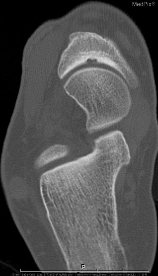
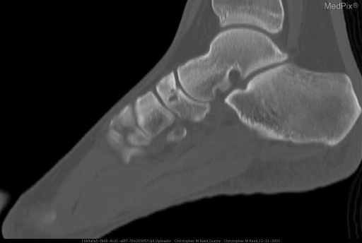
Osteochondritis Dissecans of the Tarsal Navicular
- Image ID
- MPX1936_synpic18349
- Case U_id
- MPX1936
- Modality
- CT · CT - noncontrast
- Plane
- Axial
- Location
- Musculoskeletal (Spine and Muscles)
- Age / Sex
- 40 / female
- Caption
- Cortical disruption at the proximal articular surface of the tarsal navicular bone. Multiloculated subchondral cyst adjacent to the cortical defect is noted.
- ACR Codes
- 4.4
Clinical case
- History
- 40 year old female with chronic right dorsal mid foot pain. Patient’s symptoms were relieved with therapeutic injection of the talonavicular joint.
- Exam
- N / A
- Findings
- Axial CT with sagittal reformats through the ankle reveals cortical disruption at the proximal articular surface of the tarsal navicular bone. Multiloculated subchondral cyst adjacent to the cortical defect is noted.
- Differential Diagnosis
- Stress fracture (Expect to see partial or complete sagittal fracture line through navicular) Osteonecrosis (Usually see sclerosis and/or collapse of the navicular)
- Case Diagnosis
- Osteochondritis Dissecans of the Tarsal Navicular
- Diagnosis By
- Clinical Course / Symptoms Recovery
Topic
- Category
- Trauma
- ACR Code
- -1.-1
Disease discussion
Osteochondritis dissecans is the traditional term used to describe osteochondral fracture. The osteochondral fragment may remain in situ or partially or completely detached from its origin, forming a loose body. Osteochondritis dissecans has been described in a variety of bones. The ankle accounts for 4% of all cases of OCD and the tarsal navicular bone is rarely involved. Most commonly, ankle OCD’s are seen in the talar dome.