Image
Diagnosis: Thinning and irregularity of the anterior talofibular ligament consistent with previous inversion injury.
- Image ID
- MPX1923_synpic23831
- Case U_id
- MPX1923
- Modality
- CT · CT - noncontrast
- Plane
- Axial
- Location
- Musculoskeletal (Spine and Muscles)
- Age / Sex
- 25 / male
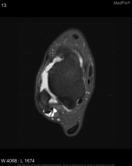
- Caption
- Axial fat-saturated T1-weighted MR image of the right ankle shows a thinned and irregular anterior talofibular ligament and fluid communicating between the joint space and the peroneal tendon sheath. No other ligamentous or tendinous abnormality, fracture or soft tissue swelling is present.
- ACR Codes
- 4.4
Clinical case
- History
- History (can include gestational age, or age in days, weeks, months): 25 year-old male with multiple right- sided inversion injuries over past ten years. Currently complains of subluxing peroneal tendons.
- Exam
- Physical Exam and Laboratory: Well-developed, white man who has no swelling, erythema, scars or point tenderness of the right ankle. He has full and equal range of motion bilaterally. He is able to sublux his peroneal tendons. Negative anterior drawer sign. No subtalar instability onphyscial examination.
- Findings
- Image Findings: Axial fat-saturated T1-weighted MR image of the right ankle shows a thinned and irregular anterior talofibular ligament and fluid communicating between the joint space and the peroneal tendon sheath. No other ligamentous or tendinous abnormality, fracture or soft tissue swelling is present.
- Differential Diagnosis
- Differential Diagnosis for these findings in this case: Chronic injury to anterior talofibular ligament.
- Case Diagnosis
- Diagnosis: Thinning and irregularity of the anterior talofibular ligament consistent with previous inversion injury.
Topic
- Category
- Trauma
- ACR Code
- 4.4
Disease discussion
Discussion (include references):
Inversion injuries are very common in the young active population. This injury may result in a fracture, ligamentous injury, or both. The initial evaluation of a patient with inversion injuries requires a good history and physical examination as well as plain radiographs to assess for any fractures. Stress views of the both ankles should show similar tibiotalar tilt (less than 5 degrees difference between the affected and normal ankle). Finally MRI has been shown as a good modality for evaluating the ligamentous structures of the lateral ankle.
The lateral ankle is supported by three ligaments, the anterior talofibular, the calcaneofibular, and the posterior talofibular ligaments. In this case the patient had thinning of the anterior talofibular ligament. This is often the first ligament damaged in an inversion injury and is seen in approximately 40% of these injuries. With increasing force the next ligament injured in an inversion injury is the calcaneofibular ligament. The final ligament injured is often the posterior talofibular ligament. Following injury as many as 20% may experience symptomatic ankle instability.
The best visualization of the ATFL and PTFL occurs on the axial image at the talar neck, while evaluation of the calcaneofibular ligament may require an oblique view. A complete interruption of the low intensity ligament is seen in a rupture of the ligament. In this case there is thinning and irregularity of the ligament.
Reference: 1) Physical Examination of the Spine and Extremities; Stanley Hoppenfeld, pp222, Prentice Hall NJ 1976
2) Netter’s Concise Atlas of Orthopaedic Anatomy; Jon C. Thompson MD, pp 254
3) Wheeless’ Textbook of Orthopaedics Online; www.wheelessonline.com
4) Ahmad, M.A. “Magnetic Resonance Imaging of the normal and injured lateral collateral ligaments of the ankle” Ann Chir Gynaecol 1998; 87(4):311-6
5)Colville, M.R., “Surgical Treatment of the Unstable Ankle”, JAAOS Vol 6, No.6