Image
Heterotopic ossification
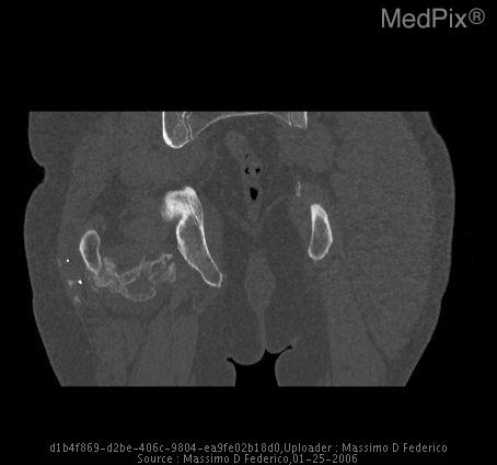
- Image ID
- MPX1905_synpic28196
- Case U_id
- MPX1905
- Modality
- CT · CT - noncontrast
- Plane
- NOS - Not specified
- Location
- Musculoskeletal (Spine and Muscles)
- Age / Sex
- 39 / male
- Caption
- A calcified mass is present posterior to the acetabulum and femoral neck.
- ACR Codes
- 4.9
Clinical case
- History
- A 39 year-old man was injured in a gunfight. The patient suffered a GSW to the right buttock that exited the scrotum. The patient underwent an exploratory laparotomy, found to be negative, and a transscrotal exploration that found injury to the bladder and urethra. A probing of the right hip wound revealed that the injury went posterior to the femur and did not invade the capsule. Approximately four months post-injury, the patient reported a progressive limiting of his hip range of motion and inability to perform activities of daily living.
- Exam
- The patient was found to have an antalgic gait. He had no neurologic symptoms and had limited mobility secondary to a mass on his right hip. His right hip range of motion was from 90 degrees of flexion to 7 degrees of flexion. His external rotation was approximately 15 degrees, and internal rotation was 30 degrees.
- Findings
- Both CT and x-ray show a calcified mass posterior to the acetabulum and femur.
- Differential Diagnosis
- Heterotopic ossification Soft-tissue neoplasm Bone neoplasm
- Case Diagnosis
- Heterotopic ossification
- Diagnosis By
- Surgery
Topic
- Category
- Trauma
- ACR Code
- 4.9
Disease discussion
Heterotopic ossification (HO) is the pathological formation of new bone in soft tissues. HO has been extensively studied and reported as a complication that develops following brain trauma, spinal cord injury, thermal injury, local joint trauma, acetabular fracture, lateral hip soft tissue hematoma, and total hip arthroplasty.(1-4) Post- traumatic ectopic calcification following a direct blow to muscle (particularly in the anterior thigh) has been described as well.(5) All forms of HO, however, are histologically identical.(4)
The incidence of such HO has been described as occurring from 5% to 90% of the time with the various traumas/surgeries.(3) Most cases of heterotopic ossification are asymptomatic, but 2% to 10% of HO can be extensive.(4)
The most common clinical manifestations of hip HO are decreased range of motion, pain around the joint, and difficulty walking.(3, 6-7) Prophylaxis recommended for HO includes nonsteroidal anti-inflammatory drugs and low dose local radiation.(8)
Once HO becomes established and extensive with joint movement restriction, the only effective treatment is surgical resection.(4)