Інші зображення цього випадку
Image
Patellar Tendinosis (Jumper's Knee) with probable partial tear of the patellar tendon.
- Image ID
- MPX1861_synpic20765
- Case U_id
- MPX1861
- Modality
- MR · MR - PDW Proton Density
- Plane
- Sagittal
- Location
- Musculoskeletal (Spine and Muscles)
- Age / Sex
- 23 / male
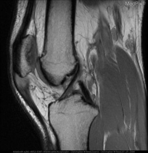
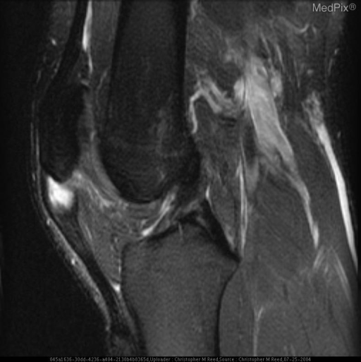
- Caption
- Sagittal Proton Density MR Image demonstrates increased signal and thickening of the patellar enthesis of the patellar tendon.
- ACR Codes
- 4.4
Clinical case
- History
- 23 year old male with acute onset left knee pain, worse with full extension.
- Exam
- No obvious external deformity. Tender to Palpation over the Patella. Relatively Weak Left Knee Extension.
- Findings
- Increased signal intensity and thickening within the patellar enthesis of the patellar tendon on Proton Density and T2 weighted sequences.
- Differential Diagnosis
- Patellar Tendinosis (Jumper's Knee) Patellar Tendon Rupture
- Case Diagnosis
- Patellar Tendinosis (Jumper's Knee) with probable partial tear of the patellar tendon.
- Diagnosis By
- MRI
Topic
- Category
- Trauma
- ACR Code
- 4.4
Disease discussion
Patellar tendinosis, also known as Jumper's Knee, is the result of micro and partial macro-tearing of patellar tendon fibers, usually due to repetitive running and/or jumping (Basketball, Football, and Volleyball). Either the patellar or tibial enthesis may be involved; however the condition is most often associated with patellar attachment involvement. Treatment is generally conservative.