Image
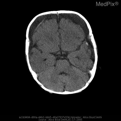
Battered child syndrome. Chronic bilateral subdural hematomas.
- Image ID
- MPX1770_synpic23443
- Case U_id
- MPX1770
- Modality
- CT · CT - noncontrast
- Plane
- Axial
- Location
- Brain and Neuro (Head)
- Age / Sex
- 1 / female
- Caption
- Axial non-contrast CT of the head demonstrates bilateral low density chronic subdural fluid collections, along with a focal high density adjacent to the left temporal lobe suggestive of more recent subdural hemorrhage.
- ACR Codes
- 1.-1
Clinical case
- History
- 5 month old female brought to ER with lethargy. Imaging request stated "hypoglycemia."
- Findings
- Non-contrast CT demonstrated bilateral subdural fluid collections
- Differential Diagnosis
- Non-accidental trauma
- Case Diagnosis
- Battered child syndrome. Chronic bilateral subdural hematomas.
Topic
- Category
- Trauma
- ACR Code
- 1.-1
Disease discussion
The spectrum of intracranial injuries in physically abused children includes: acute subdural hematoma, chronic subdural hematoma, acute epidural hematoma, cerebral contusion, focal/multifocal/diffuse cerebral edema, and atrophy. Acute and/or chronic subdural hematomas and cerebral contusion are common in child abuse. Intracerebral hematomas and epidural hematomas are uncommon.
Untreated acute subdural hematomas undergo a series of predictable pathologic changes. Soon after the acute hemorrhage, the blood collection becomes surrounded by endothelial cells and there is invasion by granulation tissue. Progressive liquefaction of the hematoma results in its conversion to a serous fluid. These serous subdural collections are referred to as chronic subdural hematomas or subdural hygromas. Chronic subdural hematomas, related to prior trauma, are most often located adjacent to one or both cerebral convexities but may also extend into the interhemispheric fissure.
The CT appearance of a subdural hematoma varies according to the time period since the injury. The initial hemorrhage is visualized as a high-attenuation fluid collection on CT. The attenuation values of the lesion progressively decrease over the next weeks to months. Chronic subdural hematomas usually contain serous fluid and produce attenuation values slightly greater than those of CSF. It is not uncommon for an abused child to have suffered multiple episodes of trauma, resulting in intracranial hematomas in different stages of evolution.
At times, chronic subdural hematomas are difficult to differentiate from enlargement of the subarachnoid spaces. In many cases, a distinct transition is visible between the subdural fluid collection and the slightly lower attenuation CSF of the subarachnoid space. A membrane of variable thickness separating the two spaces may be identifiable. The brain adjacent to a chronic subdural hematoma is often displaced, and the sulci may be compressed and effaced. In contradistinction, enlarged subarachnoid spaces are associated with interdigitation of the fluid into the cerebral sulci.
The differential diagnosis of isolated chronic subdural fluid collections in infants includes bacterial meningitis, remote trauma, rickets, and Menke's syndrome. In the absence of a clear-cut, suitable clinical history of other forms of trauma, the CT demonstration of coexistent acute and chronic subdural hematomas is highly suggestive of child abuse.