Image
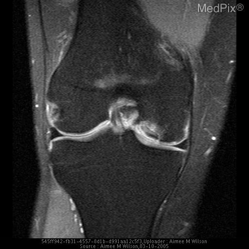
Osteochondritis Dissecans Stage IV
- Image ID
- MPX1751_synpic24225
- Case U_id
- MPX1751
- Modality
- MR · MR - T2 weighted
- Plane
- Coronal
- Location
- Musculoskeletal (Spine and Muscles)
- Age / Sex
- 22 / male
- Caption
- High signal intensity between osseous bone fragment and femoral condyle.
- ACR Codes
- 4.4
Clinical case
- History
- Hx: 22 y.o. male no chief complaint or physical exam available.
- Findings
- MRI: Intraarticular osseous fragment within the lateral aspect of medial femoral condyle with fluid seen surrounding the lesion and disruption of the articular cartilage, and slight posterior diplacement of the osseous fragment
- Differential Diagnosis
- DDx:Osteochondral fracture, chondral injuries, osteonecrosis, accessory ossicle
- Case Diagnosis
- Osteochondritis Dissecans Stage IV
Topic
- Category
- Trauma
- ACR Code
- 4.4
Disease discussion
OCD's pathologic mechanism is uncertain, so there remains no one clear etiology. A multitude of theories have developed to explain etiology, from repetitive microtrauma, and stress fractures to avascular necrosis. The most common site is in the knee. 85% of knee OCD lesions are located in medial condyle which is theorized to be caused by possible enlargement of tibial spines in combination with unusual stress against these surfaces.
MRI has utility for detecting and characterizing OCD lesions. It accurately measures lesion size and provides cartilage/subchondral bone status. The higher signal on T2 weighted sequences can show marrow changes consistent with bony edema. Interestingly, unstable OCD lesions that fail non-operative managment are most often lesions that have a high T2 signal linearity behind the OCD lesion and underlying bone. In addition, the high signal line may be consistent with a disruption of the articular cartilage seen on T1 sequences.
OCD lesion staging classification on MRI:
Stage I: small change of signal without clear margins of fragment.
Stage II: Osteochondral fragment with clear margins without fluid between fragment and underlying bone.
Stage III: Fluid is partially visible between fragment and underlying bone.
Stage IV: Fluid completely surrounds the fragment but the fragment is in situ.
Stage V: Fragment is completely detached and displaced (loose body).
Treatment/Management:
Non-operative management is based on lesion size and patient maturity. The smaller the lesion and acquiring the lesion prior to growth plate closure provides good prognostic indicators for favorable healing. Typically, with a period of immobilization and gradual weight-bearing, lesions heal. MRI can assess healing.
Operative management is considered when lesions are detached or unstable and in patients approaching physeal closure and/or non-operative treatment has failed. There are many techniques from drilling into the OCD lesion to create revascularization and healing with fibrocartilage (typically in 4-5mths), to staples, and arthrotomy with bone graph and K-wire fixation, in addition using autologous chondrocyte implantation.