Інші зображення цього випадку
Image
Quadriceps tendon rupture
- Image ID
- MPX1737_synpic52863
- Case U_id
- MPX1737
- Modality
- MR · MR - T2 weighted
- Plane
- Sagittal
- Location
- Musculoskeletal (Spine and Muscles)
- Age / Sex
- 52 / male
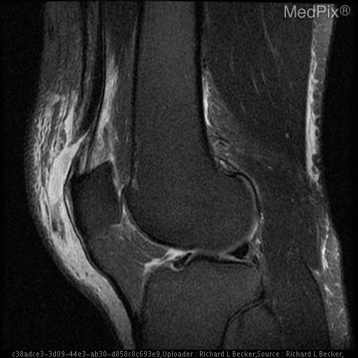
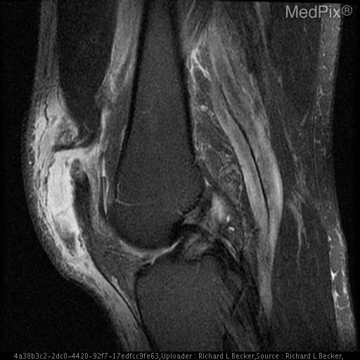
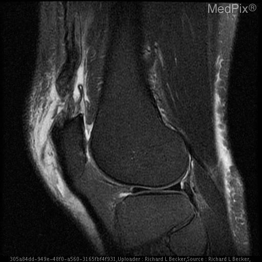
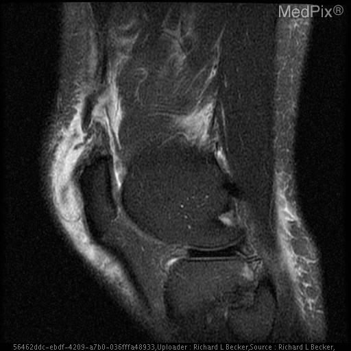
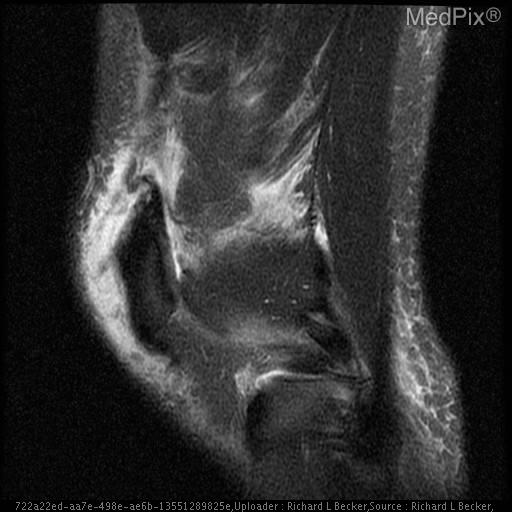
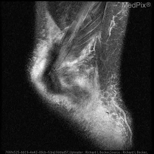
- Caption
- Joint effusion
- ACR Codes
- 4.4
Clinical case
- History
- 52 y/o male with swollen painful knee and weakness in extension s/p fall
- Exam
- Marked swelling superior aspect of joint. Mildly ballotable patella. 3/5 quad strength in extension.
- Findings
- Xray remarkable for effusion. MRI reveals complete quadriceps tendon discontinuity medially, with retraction. Some intact deep fibers laterally. Effusion and edema. Superior patellar enthesophyte. No patella alta.
- Differential Diagnosis
- Quadriceps tendon rupture
- Case Diagnosis
- Quadriceps tendon rupture
- Diagnosis By
- Patient taken to surgery for repair
Topic
- Category
- Trauma
- ACR Code
- 4.4
Disease discussion
Quadriceps tendon ruptures usually result from a rapid, strong eccentric contraction of the quadriceps muscle with the knee partially flexed, typically during a fall. This injury may also be seen after a direct blow to the quadriceps tendon. Ruptures are also known to occur spontaneously or after relatively trivial trauma, particulary in those with chronic conditions such as diabetes, chonic renal disease, rheumatoid arthritis, or any condition requiring the use of long-term steroids. These tears most commonly occur at the tendinous insertion of the quadriceps to superior pole of the patella.
Patients usually present with knee pain, swelling and difficulty ambulating. Physical exam with complete tears classically demonstrates suprapatellar swelling, a palpable defect in the suprapatellar region with tenderness, a low-riding patella, and decreased active range of motion and strength of knee extension. Partial tears are often much more difficult to discern clinically and as such may be easily misdiagnosed as a "knee sprain."
Imaging modalites such as plain radiography and MRI are often used to support the clinical findings. Findings that suggest quadriceps tear on plain radiographs include suprapatellar soft tissue swelling and distortion of the typical fat planes, and patella baja (inferiorly positioned patella). Due to the fact the injury commonly occurs at the tendinous insertion site at the patella, avulsion fractures of the patella may also be seen. MRI demonstrates disruption of the normal low intensity quadriceps tendon on T2 weighted images, which is replaced by high intensity fluid signal secondary to inflammatory response and edema. Ultrasound is another modality that may be used to demonstrate quadriceps tendon tears. However, unlike MRI, ultrasound will not demonstrate secondary associated findings often seen in traumatic injuries to the knee such as ligamentous and osteochondral insults, and as such is used much less frequently.
Partial tears are treated conservatively, typically with a 1-2 month peroid of immobilization in full knee extesion, followed by a course of physical therapy to restore range of motion and strength. Complete tears are usually treated with early surgical intervention which again is followed by a course of immobilization and physical therapy.