Інші зображення цього випадку
Image
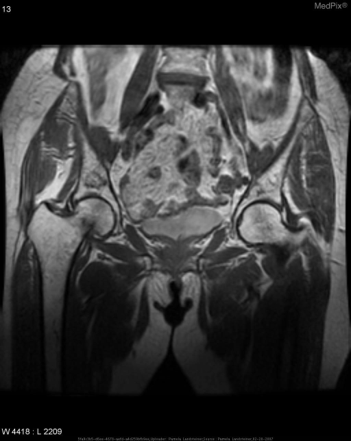
Non-displaced transverse fracture of right femoral neck.
- Image ID
- MPX1559_synpic34909
- Case U_id
- MPX1559
- Modality
- MR · MR - T1W - noncontrast
- Plane
- Coronal
- Location
- Musculoskeletal (Spine and Muscles)
- Age / Sex
- 76 / female
- Caption
- Coronal T1-weighted MR image shows linear low-signal-intensity area extending from medial cortex into the medullary canal of right femoral neck, consistent with fracture.
- ACR Codes
- 4.4
Clinical case
- History
- A 76-year-old woman presents to her primary care manager with right hip pain. No known history of trauma.
- Exam
- Physical exam information is not available.
- Findings
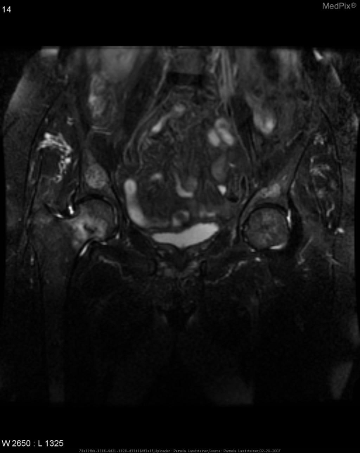
- Anterior-posterior radiograph of the pelvis, and frog-leg radiograph of the right hip show diffusely diminished bone density of the pelvis and right hip. No periosteal reaction or fracture is seen. Coronal T1-weighted MR image of the pelvis demonstrates linear low-signal-intensity area extending from the medial cortex of the right femoral neck into the medullary canal and is consistent with fracture. On the T2-weighted fat-saturated coronal MR image, more extensive area of high-signal-intensity is present consistent with a combination of the fracture and post-traumatic edema and inflammation.
- Differential Diagnosis
- Non-displaced transverse fracture of right femoral neck • Stress fracture (abnormal stress to normal bone) vs. • Insufficiency fracture (normal stress to abnormal bone)
- Case Diagnosis
- Non-displaced transverse fracture of right femoral neck.
- Diagnosis By
- Confirmed by MRI.
Topic
- Category
- Trauma
- ACR Code
- 4.4
Disease discussion
Patient had complete fracture of the R femoral neck with posterior rotation of the femoral head. Should have heightened suspicion in any elderly patient with hip pain even when there are negative radiographs. MRI or bone scan can be done to look for occult femoral neck fractures when suspicion is high. MRI is most sensitive at time of presentation. Age related bone loss is believed to be the most important factor in determining the incidence of femoral neck fractures. Fractures of the femoral neck occur primarily from low-energy injuries in the elderly and high-energy injuries in younger patients.