Інші зображення цього випадку
Image
Salter-Harris Fracture
- Image ID
- MPX1537_synpic39394
- Case U_id
- MPX1537
- Modality
- MR · MR - T2 weighted
- Plane
- Coronal
- Location
- Musculoskeletal (Spine and Muscles)
- Age / Sex
- 10 / male
- Caption
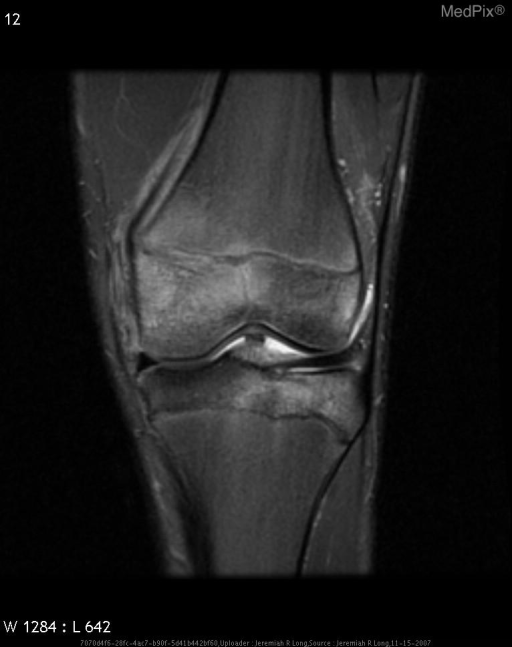
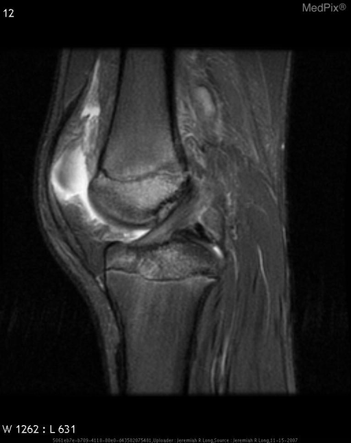
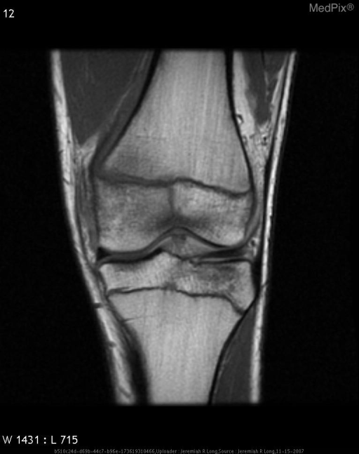
- Sagital and coronal T2 fat suppressed images demonstrate diffuse high T2 signal in the bone marrow consistent with edema in the distal femoral metaphysis and proximal tibial plateau. Additionally on the coronal T1 sequences there is an abnormal linear region of low signal extending from the joint space to the epiphysis at the intertrochanteric region of the femur. Additionally there is slight assymetric widening of the physis medially. These findings are consistent with a Salter Harris type III fracture of the distal femur.
- ACR Codes
- 4.4
Clinical case
- History
- 10 year old boy status post lateral blow while playing football.
- Exam
- N/A
- Findings
- PA and lateral radiographs of the left knee demonstrate an abnormal linear thin sclerotic line at the medial aspect of the intertorchanteric portion of the left femur as well as a joint effusion. Sagital and coronal T2 fat suppressed images demonstrate diffuse high T2 signal in the bone marrow consistent with edema in the distal femoral metaphysis and proximal tibial plateau. Additionally on the coronal T1 sequences there is an abnormal linear region of low signal extending from the joint space to the epiphysis at the intertrochanteric region of the femur. Additionally there is slight assymetric widening of the physis medially. These findings are consistent with a Salter Harris type III fracture of the distal femur.
- Differential Diagnosis
- Salter Harris type III fracture
- Case Diagnosis
- Salter-Harris Fracture
- Diagnosis By
- MRI
Topic
- Category
- Trauma
- ACR Code
- 4.4
Disease discussion
In pediatric patients, fractures through the physis represent a serious clinical concern as they can lead to potentially poor outcomes if they are not accurately diagnosed and treated appropriately. The Salter Harris radiologic classification system of physeal fractures is the most commonly utilized method of describing these fractures. This system divides fractures into five types depending upon the involvement of the physis, epiphysis, or metaphysis.
Type I fractures are transverse through the hypertrophic zone of the physis and results in widening of the physis. In these injuries, the growing zone of the physis is typically not disturbed and overall growth disturbance is uncommon.
Type II fractures traverse the metaphysis and physis, but do not involve the epiphysis. This represents the most common type of physeal fracture, accounting for up to 75% of injuries. While these may result in mild limb foreshortening, functional limitations are rare.
Type III fractures traverse the physis and the epiphysis, but spare the metaphysis. These represent a more serious clinical issue, as they tend to involve the articular cartilage and have a greater predisposition for growth arrest.
Type IV fractures involve the epiphysis, physis, and metaphysis. Like the type III injury, these fractures have a greater predisposition for growth arrest.
Type V fractures result from a crush injury to all or part of the physis. Initial diagnosis may be dificult radiographically without specific clinical data. Often the diagnosis is made in follow up, after partial or complete boney fusion across the physeal plate has ensued.
While it has been estimated that nearly 30% of these fractures result in growth plate disturbance, only 2% result in significant functional disability.