Інші зображення цього випадку
Image
ACL Tear with lateral femoral notch sign
- Image ID
- MPX1500_synpic19061
- Case U_id
- MPX1500
- Modality
- MR · MR - PDW Proton Density
- Plane
- Lateral
- Location
- Musculoskeletal (Spine and Muscles)
- Age / Sex
- 0 / N/A
- Caption
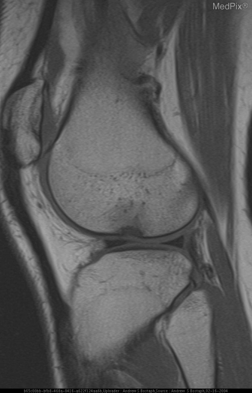
- Figure 2: Sagital PD shows a “low lying” ACL consistent with tear.
- ACR Codes
- 4.4
Clinical case
- History
- 18 y/o female with history of chronic pain with exercise. No history of specific trauma.
- Exam
- Noncontributory
- Findings
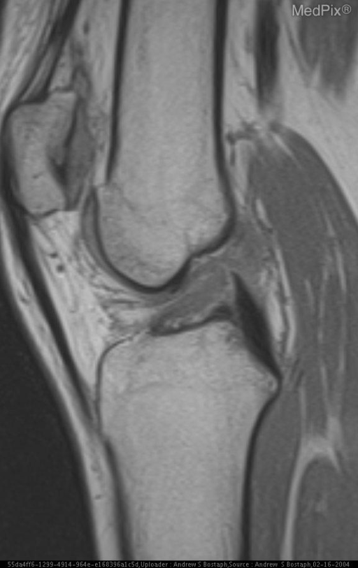
- Figure 1: Lateral radiograph in an 18 year old female with chronic pain during exercise. Figure 2: Sagital PD shows a “low lying” ACL consistent with tear. Figure 3: Sagital PD with FS shows an ostoeochondral defect of the lateral femoral condyle corresponding to the radiographic deep lateral femoral notch.
- Differential Diagnosis
- Normal Variant Osteochondral defect, nontraumatic vs traumatic
- Case Diagnosis
- ACL Tear with lateral femoral notch sign
- Diagnosis By
- MRI
Topic
- Category
- Trauma
- ACR Code
- 4.4
Disease discussion
The lateral femoral notch (lateral condylopatellar sulcus) is normally a shallow groove in the mid lateral femoral condyle. The groove is formed by the junction of the tibiofemoral and patellofemoral curvature. The sulcus is typically less than 2mm in depth and is more conspicuous than the medial sulcus due to its parallel alignment with the x-ray. The depth of the lateral sulcus can be measured by drawing a line tangential to the sulcus on the articular surface. The “lateral femoral notch sign” is a result of anterior subluxation of the tibia with impaction of the lateral femoral condyle on the posterolateral tibial plateau. This pattern of injury is described as “kissing contusions”.
The finding of a deep lateral femoral notch has been associated with anterior cruciate ligament tears. Cobby et al found that a sulcus 1.5mm or deeper is a reliable indirect sign of a torn ACL. However, this sign was only present in 12% of patients with ACL tear. Garth et al also found a statistically significant association with lateral meniscus tears as well, in particular the anterior horn.
While the lateral femoral notch sign occurs infrequently with ACL tears, careful attention to this area on the lateral radiograph of the knee can be a reliable indicator of internal deraingement of the knee.