Інші зображення цього випадку
Image
Pneumomediastinum from Blast Trauma
- Image ID
- MPX1398_synpic27377
- Case U_id
- MPX1398
- Modality
- CT · CT - GI & IV Contrast
- Plane
- Axial
- Location
- Chest, Pulmonary (Thorax)
- Age / Sex
- 24 / male
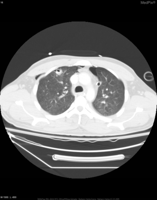
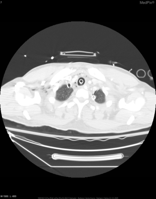
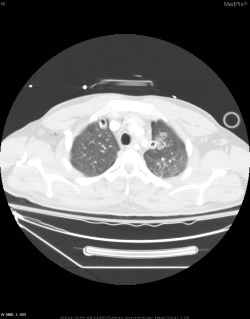
- Caption
- CT with IV and enteric contrast showing pneumomediastinum, bilateral pneumothoraces, and air in subcutaneous tissues.
- ACR Codes
- 6.7
Clinical case
- History
- 24 yo man with IED blast injury from OIF with penetrating brain injury. He underwent a decompressive right hemicraniectomy at the 10th CSH. CT chest showed bilateral pneumothoraces and pneumomediastinum, therefore bilateral chest tubes were placed. Patient was then transported to LRAMC, and finally NNMC for definitive care.
- Exam
- BP 158/65, P 91, intubation O2sats 100% GCS 3 Bilateral chest tubes and bilateral breath sounds on lung exam.
- Findings
- Pneumomediastinum with air in the anterior aspect tracking up to the thoracic inlet. Subcutaneous air noted adjacent to the chest tubes and running up into the axilla bilaterally. Pneumothoraces bilaterally. Left subclavian line, endotracheal tube, enteric tube, and bilateral chest tubes appropriately placed.
- Differential Diagnosis
- Barotrauma Penetrating injury to neck or chest positive pressure ventilation esophageal rupture postoperative mediastinitis
- Case Diagnosis
- Pneumomediastinum from Blast Trauma
- Diagnosis By
- CT with IV and enteric contrast
Topic
- Category
- Trauma
- ACR Code
- 6.735
Disease discussion
Pneumomediastinum refers to the presence of extraluminal gas within the mediastinum. This may result from alveolar rupture, perforation of the tracheobronchial tree or esophagus, or gas leaking into the thorax from the neck, chest wall, or abdomen.
Radiographically, pneumomediastinum is characterized by lucent streaks in the mediastinal soft tissues. A number of radiographic signs of pneumomediastinum result from the gas outlining normal anatomic structures.
These signs include:
thymic sail sign- elevation of the thymus
Nacleiros V sign - gas outlining the descending aorta laterally with extension laterally between the parietal pleura and medial hemi diaphragm
double bronchial wall sign- gas next to a major bronchus, delineating the bronchial walls
continuous diaphragm sign- gas extending posterior to the pericardium delineating the central portion of the diaphragm on the frontal projection
"ring around the artery" sign- gas surrounding the right main pulmonary artery as seen on the lateral radiograph
tubular artery sign- gas adjacent to the major branches of the aorta, delineating the walls medially, while aerated lung delineates the walls laterally
extrapleural sign- gas from the mediastinum extending laterally between the parietal pleura and the diaphragm