Інші зображення цього випадку
Image
Partial Achilles Tendon tear
- Image ID
- MPX1394_synpic22908
- Case U_id
- MPX1394
- Modality
- MR · MR - T1W - noncontrast
- Plane
- Lateral
- Location
- Musculoskeletal (Spine and Muscles)
- Age / Sex
- 65 / male
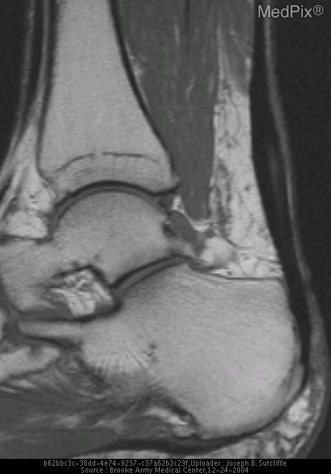
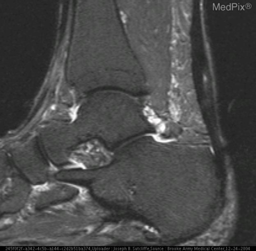
- Caption
- T1W image demonstrates focal thickening and intermediate signal within the otherwise dark signal of the Achilles tendon
- ACR Codes
- 4.4
Clinical case
- History
- Left ankle/leg pain and weakness
- Exam
- Weakness on plantar flexion
- Findings
- Plain radiographs demonstrate thickening of the Achilles tendon, convex impression of Achilles tendon on the posterior margin of Kager's fat pad, and a lower limits normal Toygar's angle. MR findings demonstrates focal thickening of the Achilles tendon increased T2W signal within the tendon and overlying subcutaneous tissues
- Differential Diagnosis
- Partial Achilles rupture Complete Achilles rupture
- Case Diagnosis
- Partial Achilles Tendon tear
- Diagnosis By
- Imaging findings were confirmatory
Topic
- Category
- Trauma
- ACR Code
- 4.4
Disease discussion
Spontaneous Achilles tendon rupture or partial tears typically occur in otherwise healthy, relatively young patients, with no history of heel or calf pathology. Most studies find that the majority of individuals who sustain Achilles tendon injury are men in their third to fifth decade of life, who are participating in sports activities. The injury most commonly occurs secondary to overloading of the musculotendinous unit in a poorly conditioned individual rather than secondary to an underlying tendinopathy. The mechanical cause of the injury is most commonly active, forceful plantarflexion, usually associated with pushing off athletic movements. Less commonly, Achilles rupture is caused by violent unexpected dorsiflexion of a plantarflexed foot, such as when a person steps in hole or falls from a height.(1)
The diagnsosis can often be made by physical examination. Findings may include a palpable depression over the area of tendon rupture, weakness of plantarflexion, and positive Thompson test. A positive Thompson test is failure of the foot to plantarflex when the calf muscles are squeezed.(1)
However, up to 25% of patients with partial or complete Achilles tear may be misdiagnosed based on physical exam findings alone. Radiographic findings can assist with the diagnsosis. When the Achilles tendon is ruptured, the sharp posterior contour of Kager's fat pad triangle will become serrated and indistinct. The Achilles tendon will appear thickened (> 8mm). Toygar's angle, which is the angle of the posterior skin surface overlying the distal Achilles and posterior calcaneal surfaces will decrease and is considered abnormal if less than 150 degrees. Positive Arner's sign, in which the anterior contour of the Achilles tendon at the insertion curves away from the superior-posterior aspect of the calcaneus, may be positive.(2)
MR imaging findings may confirm the diagnosis. Partial Achilles tendon tears demonstrate heterogeneous signal intensity and thickening of the injured tendon without complete interruption. Edema manifested as increased T2 signal will usually be present within the tendon, subcutaneous tissues, and in Kager's fat pad. Hemorrhage signal may also be present in those structures and signal characteristics will vary according to the age of the injury. Complete Achilles rupture manifests as discontinuity of the tendon with fraying and retraction of the torn edges. In acute ruptures, the gap between the rupture will have intermediate T1 signal and high T2 signal due to edema and acute hemorrhage. In chronic ruptures, scar or fat signal will ususally predominate.(3)
Treatment of Achilles rupture is controversial and there are advantages and disadvantages to conservative nonsurgical treatment versus surgical treatment. Nonsurgical treatment avoids surgical morbidity and cost, but rerupture rates are as high as 39%. Surgical treatment has higher cost and morbidity, but lower rerupture rates.(1)