Image
Osteochondritis Dissecans
- Image ID
- MPX1231_synpic27572
- Case U_id
- MPX1231
- Modality
- CT · CT - noncontrast
- Plane
- Axial
- Location
- Musculoskeletal (Spine and Muscles)
- Age / Sex
- 15 / female
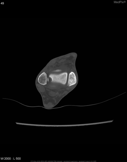
- Caption
- Noncontrast CT image of the talus demonstrates a shallow, rounded cortical defect within the superior lateral talar dome and an adjacent fragment of bone within the ankle joint.
- ACR Codes
- 4.4
Clinical case
- History
- Chronic right ankle pain with occasional locking of the joint and a history a severe right ankle injury approximately six months earlier.
- Exam
- N/A
- Findings
- Radiographs of the right ankle demonstrate a cortical lucency at the superior and lateral aspect of the talar dome. Noncontrast CT images of the talus demonstrate a shallow, rounded cortical defect within the superior lateral talar dome and an adjacent fragment of bone within the ankle joint.
- Differential Diagnosis
- Osteochondritis dessicans, normal variant ossification abnormality, acute traumatic fracture fragment.
- Case Diagnosis
- Osteochondritis Dissecans
- Diagnosis By
- CT scan.
Topic
- Category
- Trauma
- ACR Code
- 4.4
Disease discussion
Osteochondral defects are believed to be related to repeated microtrauma. They are a form of avascular necrosis that is smaller and more focal. The appearance is usually a concave subchondral fracture line containing an osseous body. It is frequently found in the knee, most commonly in the lateral portion of the medial femoral condyle. Alternatively, it is also seen in the dome of the talus and the capitellum. MRI examination can be performed to evaluate the stability of the osseous body and to check for effusion tracking along its borders in symptomatic patients. Evaluation with gadolinium show high signal intensity surrounding the fragment, this is felt to represent granulation tissue formation around an unstable fragment. Differential considerations include spontaneous osteonecrosis which usually affects older patients who complaint of severe pain and shows flattening of the weight bearing surface of the femoral condyle on radiographic examination.